Lipopolysaccharide (LPS) covers the outer surface of Gram-negative bacteria. Lipid A is the part that anchors it and the part our immune system senses. We develop tandem mass spectrometry methods that work out lipid A structures from a single colony and map the rest of the molecule, including O-antigen. We also use lipid spectra as barcodes to identify bacteria and detect colistin resistance straight from clinical samples.
Lipid A
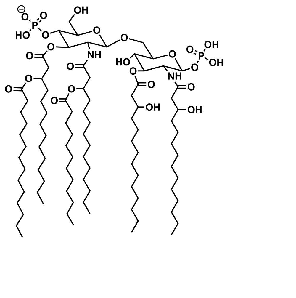
Structure
Lipid A structure from a single bacterial colony, by tandem mass spectrometry
Lipid A structural diversity in Leptospira
Abanoub Mikhael, Sophie Culos
Growing Leptospira in conditions that mimic infection, without animal models
Angela Jackson, Sophie Culos
A top-down assay for rough-type O-antigen
Structures for half of more than 500 ions in an LPS mixture, from two tandem spectra
Abanoub Mikhael
Lipid A double-bond positions mapped by ozonolysis on the MALDI plate, from a single colony
Abanoub Mikhael
Mass defect and ion mobility analysis of complex mixtures from two Gram-negative species
Abanoub Mikhael
A previously unknown structure from Pragia fontium
Linda Nartey, Abanoub Mikhael
Lipid A structural variation in Akkermansia species
Silas Porter Crenna
Lipid A features behind temperature adaptation in Francisella, and the fragmentation rules that came from them
How lipid A structure affects recognition by toll-like receptors
In the clinic
Lipid spectra as barcodes: bacteria identified directly from the specimen, with no culture
Detecting lipid changes that make bacteria resistant to antimicrobial peptides
FLAT: lipid extraction and mass spectrometry on a single MALDI spot in under an hour
Urinary tract infections identified within an hour of reaching the lab, without culture
Screening suspected urinary tract infections in a hospital lab with over 90% accuracy, against a dipstick test with over 70% false positives
Linda Nartey